NEW STUDY! Parasym Plus™ for Multiple Sclerosis › Forums › PrettyIll.com Discussion › EDS/MS/Chiari › Could someone please explain this upright MRI finding
- This topic has 15 replies, 3 voices, and was last updated 8 years, 5 months ago by
Barbara.
-
AuthorPosts
-
October 17, 2015 at 9:02 am #904
misosoup
ParticipantHello
I had an upright MRI recently which showed many things, including retroflexed odontoid and some subluxation.
I was sent for the MRI as the EDS specialist I saw suspected I had a Chiari, as I have many symptoms, including a brain test that showed issues with my cerebellum/ brainstem.
This is what part of the report says-
‘The cerebellar tonsils lie lower than normal, and just extending through the foramen magnum.
This degree of cerebellar tonsillar ectopia is within the normal range, but also isn’t lower than seen in the general population’‘At the cranio- cervical junction there is a very mild degree of cerebellar tonsillar ectopia with the tonsils lying within the foramen magnum’.
The odontoid issues has been explained to me, and I’m being referred to a Neurosurgeon.
Could someone please explain what they mean about the cerebellar tonsils?
Thanks
October 21, 2015 at 8:45 am #5668Dr. Diana
KeymasterHi there! Your doctor should be able to show you your images to explain this — it would be great to see, I think. What it means (basically) is that you *almost* have a Chiari-type presentation, with the back part of your brain sort of hovering over the spot where your CSF normally drains into your spinal column. Sometimes this can cause Chiari symptoms, sometimes people are never symptomatic even when that part of the brain dips so far down that the patients have true Chiari. Knowing this, I think it is critical to rule out high intracranial pressure, which can cause/exacerbate this presentation (as can tethered cord). This is one thing that my kids are I presented with, yet were able to avoid Chiari surgery, brain shunts and neck fusions by reducing our intracranial pressure. Information is power, right? Big hug to you…
October 22, 2015 at 11:13 am #5670ParticipantThank you very much for replying!
When you say ‘almost Chiari’ would this be a Chiari O?
Does this mean that this ‘almost Chiari’ could be causing some CSF blockage?
I have the images on disc, would it be helpful to post them here for you to see?
The ‘cerebellar tonsils’ part is the only part of the report that I didn’t really understand. But thanks very much for clarifying that for me.
When I saw the EDS specialist, as soon as he saw my symptom list, he suspected a Chiari.
I have been told I have a ‘retroflexed odontoid peg’ which is causing ‘ventral brainstem compression’. I’m not sure why this has happened, but it could due to part of my neck ‘sub-luxing’ when I move it. This, by the looks of it, is definitely causing a blockage.
I am now waiting to see a Neurosurgeon.
In 2009, I was referred to Ear Nose and Throat as many of the symptoms I’d been suffering for years had become unbearable. I was having the following symptoms- pain and pressure in my head feeling like my skull was going to explode, nausea and vomiting, vertigo, dizziness, balance problems, ear fullness/ popping/ pressure, pulsatile tinnitus, tinnitus, a sense of fluid behind my ears ‘whooshing’ sound in ears, hearing problems, neck pain.
My ears were fine, and they did a bunch of tests that showed abnormal eye movements and concluded that my symptoms were neurological. (Brainstem/ cerebellum).
Unfortunately these results were not passed on to anybody, so I just got worse and worse.
A year ago I was assessed by a brain injury organisation who said I had extensive brain injury symptoms- speech, memory, cognitive, movement, balance etc. But they obviously couldn’t tell me what was the cause.
Since these findings, I contacted them again and they said that this ‘retroflexed odontoid’ would be causing a CSF blockage (going in)- let alone the pressing on the brainstem, which would explain the ‘feeling like my head is going to explode’ sensation.
They’ve also gone through a list of my symptoms- speech, memory etc, to show the Neurosurgeon to show which parts of my brain could be damaged by some kind of fluid build up.
I’ve likely had this blockage/ brainstem compression for over 10 years.
Does this make sense to you, with your experience of people with this kind of condition?
October 22, 2015 at 1:33 pm #5671KeymasterYes! This is not uncommon in our patient population, certainly. On caveat: MANY of us (myself and my children included) have this presentation (including the retroflexed odontoid, etc) and yet do not need surgery. Instead, when we lower our intracranial pressure a bit (with Diamox, usually), the brain lifts a bit, getting the offending anatomy out of the way of the brain stem and the foramen (allowing CSF to flow through again). It’s so important to see if you can reduce or eliminate symptoms (and signs) by lowering your intracranial pressure PRIO to considering surgery. Sometimes, we don’t end up needing surgery to correct this (even small Chiari can go away when the brain floats more properly). Sometimes the presentation changes enough to alter the surgery, should surgery still be needed. And sometimes, our high intracranial pressure goes away over time, making both Diamox and surgery unnecessary. Does that make sense? Thanks for sharing — great image captured! 😉
October 22, 2015 at 2:22 pm #5674ParticipantThank you very much for taking the time to answer my questions!
The specialist said about the Chiari, as you have said, that it can be managed with medication and physiotherapy, and pain management.
He was mostly concerned about the retroflexed odontoid, which is why I’m being referred to a Neurosurgeon.
Also, the ‘left facet of the atlanto axial joint’ (no idea what that means!) is subluxing when I move my head. This is likely the cause of the ‘retroflexed odontoid’.
Would you be able to recommend anything I can do in the meantime such as- supplements or exercises? I take Vitamin D, also magnesium (after watching your video), B vitamin complex.
Thanks again for taking the time to reply.
November 14, 2015 at 7:53 pm #5690Barbara
ParticipantThe atlanto-axial joint is the joint between C1 and C2, these are the top bones in your neck, so if head movements are causing subluxation here, that means you have a degree of cervical instability between these bones.
The head and neck should move as one unit, so if one bone turns, the bone below it should move correspondingly. In your case these two bones are moving independent of each other to some degree, causing problems.
More importantly, I have looked at your MRI and have annotated it below because I think you have a problem at the craniocervial junction too (between head and neck, or C0 – C1). Just looking at the distance between the bottom of your skull, at the back and the top of C1, at the back, the gap seems too wide. Have you been in an accident ?
Regards
Barbara
(UK)November 15, 2015 at 1:25 pm #5694ParticipantHi!
Can you explain more, please?
I imagine that the Neurosurgeon will spot this too?
I fell from a tree as a 10 year old, flat onto my back, from around 7 feet. That’s the only accident I’ve had, really.
Just adding another image, to see if the ‘gap’ thing is clearer?
November 15, 2015 at 7:01 pm #5695ParticipantYes the gap is definitely wide and I can see that if you’ve injured the ligaments which hold your head on, that your ‘retroflexed odontoid’ will be the ‘pivot point’ and will be pressing into your brainstem when you look down or bend down. The trouble is, you don’t feel any pain at the point where this happens, so you don’t realise what damage is being done, it just generates symptoms – maybe some immediately but they often occur some time later.
Although a low-lying cerebellum can exist without symptoms, depending upon each individuals anatomy and whether or not the CSF is obstructed, when this is combined with cranio-cervical instability, it’s a whole new picture and I believe your trouble is more likely to be hightened by instability. I’d be interested to hear what the consultant says.
see http://prettyill.com/forums/viewthread/712/
This forum topic above talks a little more about the overall possible elements involved and the mechanism, you might find it informative. At approx post 8, there’s a comprehensive summary along with possible methods of treatment and, although it does mention the Zantac and Zyrtec protocol further down, I’d try and keep this to an absolute minimum.
November 15, 2015 at 7:11 pm #5696ParticipantThanks!
Do you know what this gap means?
I’ve done some Googling, and I believe it’s the ‘atlanto-occipital gap’?
Btw, why do you ask if I’ve been in an accident? Is this not related to EDS?
I’ve had the report back from the upright MRI, and there’s no mention of this ‘gap’. How was this missed?
I’ve been looking at loads of MRIs to compare and yes, there is a definite gap there!
I do have really bad neck pain, constantly.
Thanks again
November 15, 2015 at 7:58 pm #5697Participant1. Do you know what this gap means?
2. I’ve done some Googling, and I believe it’s the ‘atlanto-occipital gap’?
3. Btw, why do you ask if I’ve been in an accident? Is this not related to EDS?
4. I’ve had the report back from the upright MRI, and there’s no mention of this ‘gap’. How was this missed?
To answer your questions:
1. The gap means that the elements between the two structures are likely being stretched
2. Yes, occipital refers to lower back of head, atlanto refers to the atlas which is C1, the top neck bone, the bone the head sits on.
3. I asked if you’d been in an accident because with EDS the gap is generally more uniform and usually less obvious.
4 Just search online for ‘missed craniocervical injuries’ and you will see that it is commonly missed!!!November 16, 2015 at 4:21 am #5698ParticipantTo answer your questions:
1. The gap means that the elements between the two structures are likely being stretched
2. Yes, occipital refers to lower back of head, atlanto refers to the atlas which is C1, the top neck bone, the bone the head sits on.
3. I asked if you’d been in an accident because with EDS the gap is generally more uniform and usually less obvious.
4 Just search online for ‘missed craniocervical injuries’ and you will see that it is commonly missed!!!I was wondering if it makes a difference as my MRI was upright?
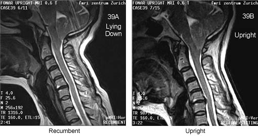
November 16, 2015 at 4:25 am #5699ParticipantI found two MRIs to compare. (The below aren’t my scans)
There’s a difference in gap size because it’s upright/ lying down.
November 16, 2015 at 7:52 pm #5700ParticipantYes there are obvious differences, I can see your head swivels forwards in the upright one, similar to what I observed at:-
http://prettyill.com/forums/viewthread/712/P30/#4183
Where, at the end, I said it looked like my hind brain (the cerebellum) was being scooped up by the back of my skull and hoiked upwards and forwards towards my brainstem.
Your scans remind me of the results of a study done, I think it was in Aberdeen, anyway I believe it was in Brain Injury 2010, (Freeman and Heffez were some of the authors involved, if you want to search on it) where they looked at the differences in the level of cerebellar tonsilar ectopia (hindbrain escaping through the bottom of the skull) between MRI’s taken laying down and upright. It was quite informative.
November 17, 2015 at 9:12 am #5701ParticipantHi Barbara
Sorry for the confusion.
Just to say the two MRIs I posted to compare (upright vs lying down), aren’t mine. They’re just some random ones I found online.
I wanted to see if an upright and lying down MRI made a difference with this gap thing. Obviously the ‘gap’ is a bit wider in the upright.
I’ve e-mailed the doctor who assessed my MRIs to ask about this gap.
November 17, 2015 at 1:17 pm #5702ParticipantHi
I just got a reply and he stated he didn’t think the gap was wide. That I have ‘laxity of the craniovertebral ligament complex and consistent with me suffering from the hypermobility syndrome’
-
AuthorPosts
- You must be logged in to reply to this topic.